How to Decide Which Antibodies to Use Immunoflouresence
It is most important that select second antibody according to the source of the primary antibody. Incubate 24 μm thick sections in three 100 mL washes of xylene for 5 minutes each for a total of 15 minutes.
Antibodies 101 Introduction To Immunofluorescence
Robert C 1 Agut H Aubin JT Collandre H Ingrand D Devillechabrolle A LeHoang P Huraux JM.

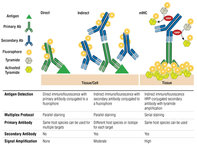
. Advances in cancer treatment using immunotherapies has accelerated the need to examine the tumor immune microenvironment TIME. See Note 3 Incubate sections in two 100 mL. Indirect IF is using two antibodies for the staining.
You can add the solution and incubate in the same way as the primary. Either different subgroups from the same species eg mouse IgG1 and IgG2a or different species. If serum contains basement zone BMZ antibodies on split-skin substrate patterns will be reported as.
Labeling and use of monoclonal antibodies in immunofluorescence. Primary antibody that specifically binds to epitope and a matched secondary antibody conjugated with fluorescence dye. Decide how many micro-liters will be needed to cover each slide.
Please use one of the following formats to cite this article in your essay paper or report. The antibody that binds to the primary antibody is called secondary antibody. For example if the primary antibody is raised in mouse the second antibody should be select as goat-anti-mouse antibody if the primary antibody is raised in.
Standard immunofluorescence staining with widefield microscopy has long been used to diagnose viral infections such as influenza from nasopharyngeal swabs from patients. The unlabeled first primary antibody specifically binds the target molecule and the secondary antibody which carries the fluorophore recognizes the primary antibody and binds to it. The successful application of mIF requires highly validated.
Using too much primary and secondary antibodies can lead to aberrant staining. Secondary antibodies can be engineered to carry different-colored fluorophores ie. 1Laboratoire de Bactériologie-Virologie CERVI Hôpital de la Pitié-Salpêtrière Paris.
1 Epidermal pattern consistent with pemphigoid. IF allows for excellent sensitivity and amplification of signal in comparison to immunohistochemistry. Most secondary antibodies can be used after 1200-1500 dilution for 1 hour at room temperature or overnight at 4 C.
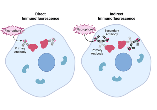
The antibody that directly binds to the antigen is called the primary antibody. Direct IF is using a single primary antibody that is conjugated with fluorescent dye. Direct IF indirect IF and combined IF.
Author Christoph R Bauer 1 Affiliation 1 Bioimaging Platform University of. A serological study of human herpesvirus-6 HHV-6 infection was performed. Detection of antibodies to human herpesvirus-6 using immunofluorescence assay.
Report includes presence and titer of circulating antibodies. Imu-no-flooo-resens a method of determining the location of antigen or antibody in a tissue section or smear using a specific antibody or antigen labeled with a fluorochrome. Multiple secondary antibodies can bind a single primary antibody.
Optimization Tips for Immunohistochemistry and Immunofluorescence. Using Antinuclear Antibody Immunofluorescence Assay. There are two major types of immunofluorescence techniques both based on the antigen--antibody reaction in which the antibody attaches itself to a specific antigen.
How to choose second antibody for IF. On the other hand. Immunofluorescence IF is an important immunochemical technique that allows for detection and localization of a wide variety of antigens in different types of tissues of various cell preparations.
Secondary indirect immunofluorescence uses two antibodies. The use of two antibodies to visualize an antigen provides the advantage of picking what color will represent the location of the antigen. Negative in normal individuals.
In addition incorparate DAPI into your secondary antibody staining. Antibody dilutions - When you are setting up staining protocols it is useful to determine the lowest dilution of primary and secondary antibodies that produce a real signal. Dilute the fluorophore-conjugated secondary antibody in PBS-005 Tween 20 again you can include 5 goat serum if the secondary antibodies are from goat.
Antibodies can recognize epitopes in their denatured linear primary form linear epitope or their native 3D tertiary form conformational epitope. Dual Labeling Using Fluorescence. Dont use a mounting media with DAPI because mounting.
Needs to determine the root cause of the positivity. Antibodies that recognize linear epitopes under denaturing and reducing conditions like in SDS-PAGE may not detect targets whose linear epitopes are concealed in the native protein structure. Guidelines for clinical use of the antinuclear antibody test and tests for.
Make sure you have the matching secondary fluorescence antibodies. Protocols for cytoskeletal and nuclear antigens Methods Mol Biol. Indirect immunofluorescence is often used to determine whether a patient is producing antibody against a particular infectious agent in order to confirm a diagnosis.
Multiplex immunofluorescence mIF assays allow the researcher to examine the spatial biology of tissues to determine tumor and immune cell interactions. Any type of immunofluorescence IF technique will require the use of a fluorescent-labeled antibody that is used to identify and located proteins antigens or other biological molecules present. An antibody against a virus-specific protein is used to stain the tissue of interest and a clinician can quickly determine whether the virus is present in the sample.
A direct fluorescent antibody DFA or dFA also known as direct immunofluorescence is an antibody that has been tagged in a direct fluorescent antibody testIts name derives from the fact that it directly tests the presence of an antigen with the tagged antibody unlike western blotting which uses an indirect method of detection where the primary antibody binds the target. 2 Dermal pattern consistent with epidermolysis bullosa acquisita. There are three types of IF.
Choose two primary antibodies with different IgG isotypes. Formalin-fixed Paraffin Embedded FFPE Slide De-paraffinization and rehydration. Place slides in 60C oven for 30 min or overnight at 37C.
For indirect immunofluorescence assay specific antibodies against the corresponding antigen fluorescein labeled anti - antibody anti - specific IgG fluorescent antibody and the primary antibody Although the basic steps and principles of immune fluorescence are the same but because of the specific conditions are not the same the detailed operation steps of each.
Learn About Immunostaining With Antibodies To Characterize Cells And Tissues Cell Signaling Technology
Direct Vs Indirect Immunofluorescence Abcam

Immunofluorescence Test An Overview Sciencedirect Topics
No comments for "How to Decide Which Antibodies to Use Immunoflouresence"
Post a Comment